PURPOSE:
To retrospectively investigate whether parenchymal enhancement in dynamic contrast material-enhanced magnetic resonance (MR) imaging of the contralateral breast in patients with unilateral invasive breast cancer is associated with therapy outcome.
MATERIALS AND METHODS:
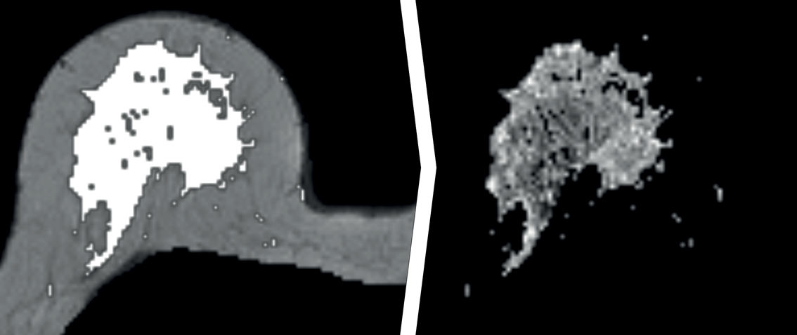
After obtaining approval of the institutional review board and patients’ written informed consent, 531 women with unilateral invasive breast cancer underwent dynamic contrast-enhanced MR imaging between 2000 and 2008. The contralateral parenchyma was segmented automatically, in which the mean of the top 10% late enhancement was calculated. Cox regression was used to test associations between parenchymal enhancement, patient and tumor characteristics, and overall survival and invasive disease-free survival. Subset analyses were performed and stratified according to immunohistochemical subtypes and type of adjuvant treatment received.
RESULTS:
Median follow-up was 86 months. Age (P < .001) and immunohistochemical subtype (P = .042) retained significance in multivariate analysis for overall survival. In patients with estrogen receptor-positive and human epidermal growth factor receptor 2 (HER2)-negative breast cancer (n = 398), age (P < .001), largest diameter on MR images (P = .049), and parenchymal enhancement (P = .011) were significant. In patients who underwent endocrine therapy (n = 174), parenchymal enhancement was the only significant covariate for overall survival and invasive disease-free survival (P < .001).
CONCLUSION:
Results suggest that parenchymal enhancement in the contralateral breast of patients with invasive unilateral breast cancer is significantly associated with long-term outcome, particularly in patients with estrogen receptor-positive, human epidermal growth factor receptor 2-negative breast cancer. Lower value of the mean top 10% enhancement of the parenchyma shows potential as a predictive biomarker for relatively poor outcome in patients who undergo endocrine therapy. These results should, however, be validated in a larger study.
