OBJECTIVES:
Ductal carcinoma in situ (DCIS) is a risk factor for incomplete resection of breast cancer. Especially, extensive DCIS (E-DCIS) orextensive intraductal component often results in positive resection margins. Detecting DCIS around breast cancer before treatment may therefore alter surgery. The purpose of this study was to develop a prediction model for E-DCIS around early–stage invasive breast cancer, using clinicohistopathological and dynamic contrast-enhanced magnetic resonance imaging (MRI) features.
MATERIALS AND METHODS:
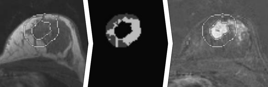
Dynamic contrast-enhanced MRI and local excision were performed in 322 patients with 326 ductal carcinomas. Tumors were segmented from dynamic contrast-enhanced MRI, followed by 3-dimensional extension of the margins with 10 mm. Amount of fibroglandular tissue (FGT) and enhancement features in these extended margins were automatically extracted from the MRI scans. Clinicohistopathological features were also obtained. Principal component analysis and multivariable logistic regression were used to develop aprediction model for E-DCIS. Discrimination and calibration were assessed, and bootstrapping was applied for internal validation.
RESULTS:
Extensive DCIS occurred in 48 (14.7%) of 326 tumors. Incomplete resection occurred in 56.3% of these E-DCIS-positive versus 9.0% of E-DCIS-negative tumors (P < 0.001). Five components with eigenvalue exceeding 1 were identified; 2 were significantly associated with E-DCIS. The first, positively associated, component expressed early and overall enhancement in the 10-mm tissue margin surrounding the MRI-visible tumor. The second, positively associated, component expressed human epidermal growth factor receptor 2 and amount of FGT around the MRI-visible tumor. The area under the curve value was 0.79 (0.76 after bootstrapping).
CONCLUSIONS:
Human epidermal growth factor receptor 2 status, early and overall enhancement in the 10-mm margin around the MRI-visible tumor, and amount of FGT in the 10 mm around the MRI-visible tumor were associated with E-DCIS.